


Metabolic imaging is a valuable noninvasive method for studying living cells with laser light, but it’s been constrained by the way light scatters when it shines into tissue, limiting the resolution and depth of penetration. MIT researchers have developed a new technique that more than doubles the usual depth limit while boosting imaging speeds, yielding richer and more detailed images.
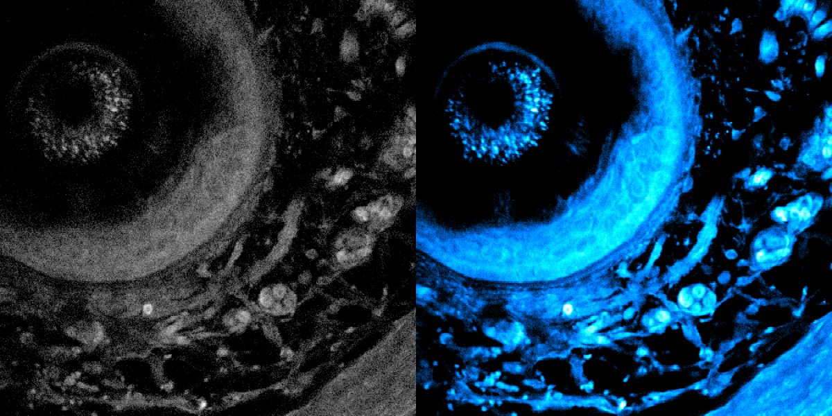
This technique does not require samples to be sliced and stained with contrast dyes. Instead, when a specialized laser shines light deep into tissues, certain molecules within them emit light of different colors, revealing molecular contents and cellular structures. By using a recently developed fiber shaper—a device controlled by bending it—the researchers can tune the color and pulses of light to minimize scattering and maximize the signal. This allows them to see much further and capture clearer images. In tests, the light was able to penetrate more than 700 micrometers into a sample, whereas the best previous techniques reached about 200 micrometers.
This method is particularly well suited for applications like cancer research, tissue engineering, drug discovery, and the study of immune responses. “It opens new avenues for studying and exploring metabolic dynamics deep in living biosystems,” says Sixian You, an assistant professor of EECS and senior author of a paper on the technique.
